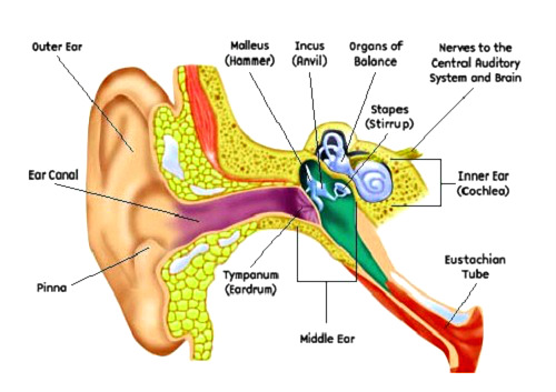

Ear Diagram below shows the various parts of a human ear. Ear anatomy includes external, middle and inner parts.
The outside portion, called the outer ear includes pinna, a ridged cartilage, through which sound travels into the short tube called the external auditory canal. This canal goes all the way to the eardrum.
Sound waves vibrate the eardrum connected to the cochlea, part of the inner ear. Sounds are then converted into nerve impulses that are further conducted to the brain. The semicircular canals are attached to the nerves and cochlea in the inner ear and are filled with fluid. Their function is to communicate the position of the head and body’s balance to the brain. The eustachian tube allows fluid to flow from the middle ear into the throat.
Essentially, a human ear is a system consisting of three organs that work together on hearing and balance in conjunction with the nervous system. The diagram below displays the human ear anatomy.