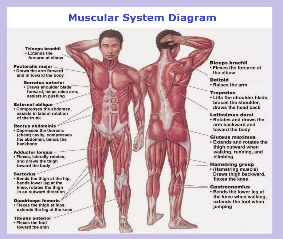
The muscular system diagram depicts the anatomy of various human muscles including biceps, tripezius, deltoid, triceps, abdominis (abs) and other key muscle groups. While multiple groups exists, overall, there are 3 different types of muscle tissues: skeletal, visceral and cardiac.
Skeletal muscles are attached to the bones, often via a joint with muscles used to bring the bones closer together. They are usually attached to the skeleton. These muscles are also controlled consciously by a human while performing such tasks as running, speaking and typing. The cells in these skeletal muscles are made of multiple progenitor cells coming together into the long and strong fibers. The diagram below summarizes skeletal muscles.
Visceral muscles are the weakest of all muscles and are a part of the organs and body systems such as blood vessels and stomach. They are used to move matter through the organ (e.g. blood, food, etc) and appear smooth and uniform when looking at the microscope. They are unconsciously controlled by the brain.
Cardiac muscle is located in the heart and pumps blood through the arteries. Similarly to visceral muscles, it is not controlled consciously; instead it stimulates itself while adjusting to the hormone levels. Cardiac tissue is very strong. Learn all about muscles.

